Shoulder

Shoulder Arthritis

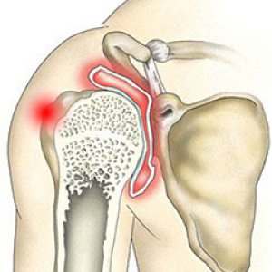
Although most people think of the shoulder as multiple joints, there are actually two joints in the shoulder area. One is located where the clavicle (collar bone) meets the tip of the shoulder bone (acromion). This is called the acromioclavicular joint or AC joint.

The junction of the upper arm bone (humerus) with the scapula (shoulder blade) is called the glenohumeral joint or scapulothoracic joint. Both joints can be affected by arthritis. To provide you with effective treatment, your doctor will have to determine which joint is affected and what type of arthritis you have.
Biceps Tendon Tear at the Shoulder

The biceps muscle in the front of your arm helps you bend your elbow and rotate your arm. It also helps keep your shoulder stable.

Tendons attach muscles to bones. Your biceps tendons attach the biceps muscle to the bones at the shoulder and at the elbow. If you tear your biceps tendon at the elbow, you will lose strength in your arm and not be able to forcefully turn your arm from palm down to palm up.

Many people can still function with a biceps tendon tear, and they only need simple treatments to relieve symptoms. Some people need surgery to repair the torn tendon.

Clavicle fracture

Posterior Sternoclavicular Dislocations

Most heal without surgery.

MIDDLE THIRD

DIAGNOSIS:

  • Pain and deformation in the middle of the bone.
  • Crepitus to movement.

Treatment:

  • Ice in the injured area.
  • Athlete’s shoulders “back” (bringing the shoulder blades or paddles together).
  • Bandage placement at “8”, elbows free.
  • You must suspend sports activity.
  • Send to X-ray.

Posterior dislocation can be life-threatening due to compression of the trachea and large blood vessels.

DIAGNOSIS:

  • Difficulty in breathing like “snoring” may occur.
  • Dysphagia (trouble passing fluid or food).
  • Changes in the color of the hand (pale or purple).
  • Arm and / or hand paresthesia (“cramps”, “burning” sensation “tingling”).

Treatment:

  • Immediate reduction (urgent).
  • Towels folded between the shoulders (“rolled up”).
  • Or shoulder hanging over the edge of the table.
  • Elbow flexed to 90 degrees, proximal arm supported.
  • Press forward.
  • If there is no immediate reduction, urgent transportation to the hospital.

Maneuver to reduce posterior sternoclavicular dislocation:

  • Upward pressure to reduce pain in collarbone.
  • Gentle longitudinal traction.

Clavicle fracture

Most heal without surgery.

MIDDLE THIRD

DIAGNOSIS:

  • Pain and deformation in the middle of the bone.
  • Crepitus to movement.

Treatment:

  • Ice in the injured area.
  • Athlete’s shoulders “back” (bringing the shoulder blades or paddles together).
  • Bandage placement at “8”, elbows free.
  • You must suspend sports activity.
  • Send to X-ray.

Posterior Sternoclavicular Dislocations

Posterior dislocation can be life-threatening due to compression of the trachea and large blood vessels.

DIAGNOSIS:

  • Difficulty in breathing like “snoring” may occur.
  • Dysphagia (trouble passing fluid or food).
  • Changes in the color of the hand (pale or purple).
  • Arm and / or hand paresthesias (“cramps”, “burning” sensation “tingling”).

Treatment:

  • Immediate reduction (urgent).
  • Towels folded between the shoulders (“rolled up”).
  • Or shoulder hanging over the edge of the table.
  • Elbow flexed to 90 degrees, proximal arm supported.
  • Press forward.
  • If there is no immediate reduction, urgent transportation to the hospital.

Maneuver to reduce posterior sternoclavicular dislocation:

  • Upward pressure to reduce pain in collarbone.
  • Gentle longitudinal traction.

Posterior dislocation can be life-threatening due to compression of the trachea and large blood vessels.

DIAGNOSIS:

  • Difficulty in breathing like “snoring” may occur.
  • Dysphagia (trouble passing fluid or food).
  • Changes in the color of the hand (pale or purple).
  • Arm and / or hand paresthesia (“cramps”, “burning” sensation “tingling”).

Treatment:

  • Immediate reduction (urgent).
  • Towels folded between the shoulders (“rolled up”).
  • Or shoulder hanging over the edge of the table.
  • Elbow flexed to 90 degrees, proximal arm supported.
  • Press forward.
  • If there is no immediate reduction, urgent transportation to the hospital.

Maneuver to reduce posterior sternoclavicular dislocation:

  • Upward pressure to reduce pain in collarbone.
  • Gentle longitudinal traction.

Thigh Hematoma

Mistreatment can lead to increased bleeding, infection, or calcification.

DIAGNOSIS:

  • Increased volume (mass) that occurs as a consequence of direct trauma.
  • Change in skin color.
  • Palpable liquid content (mainly if it is superficial).
  • If it is a “hard” consistency tumor, rule out the presence of muscle rupture.

Treatment:

  • Ice for 20 minutes, minimum.
  • Never apply heat in the acute stage of injury.
  • Place a piece of “felt”, foam rubber, a folded towel plus a compression bandage on the bruised area.
  • Do not puncture hematoma on the playing field or in the locker room.
  • If it is very extensive and close to a joint, immobilize the corresponding joint.
  • You must suspend activities.