

The knee is the largest joint in the body, and healthy knees are required for most daily activities.
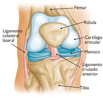
The knee is made up of the lower end of the thighbone (femur), the upper end of the shinbone (tibia), and the kneecap (patella). The ends of the three bones where they touch are covered with articular cartilage, a smooth substance that protects the bones and allows them to move easily.
The menisci are located between the femur and the tibia. These two wedge-shaped pieces of cartilage act as “shock absorbers” that cushion the joint.
Long ligaments hold the femur and tibia together and provide stability. Long thigh muscles give strength to the knee.